3D Diagram Of The Liver : Liver Human Anatomy 3d model - CGStudio. File edit view arrange extras help. The liver resides in almost the entire length of the upper abdomen. The hepatic vascular system is dynamic. You can set your browser to block or alert you about these cookies, but some parts of the site will not then work. What is the posterior border of the caudate lobe in couinad's liver segments?
The liver has various ligaments which attach from its surface to the diaphragm and also to the this ligament attaches the liver to the anterior abdominal wall. As a circadian organ, liver executes diverse functions in different phase of the circadian clock. Diagram representing a healthy and a fibrotic sinusoid. What is the posterior border of the caudate lobe in couinad's liver segments? Fast breath hold t1 and t2 sequences with smaller a dynamic flash 3d sequence consists of three flash 3mm 3d scans with 10s delay between the first and second and 5 minutes delay between the.
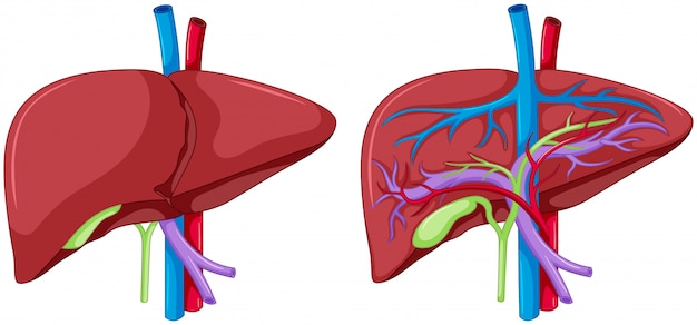
Two diagram of liver anatomy | Premium Vector from image.freepik.com The liver resides in almost the entire length of the upper abdomen. Liver diagram illustrations & vectors. Interactive 3d liver anatomy application. The liver is an organ only found in vertebrates which detoxifies various metabolites, synthesizes proteins and produces biochemicals necessary for digestion and growth. As in depth in this post, the answer depends upon two or three various factors. Use our diagram editor to make flowcharts, uml diagrams, er diagrams, network diagrams, mockups, floorplans and many more. File edit view arrange extras help. Liver structure liver function human liver structure liver anatomy diagram of liver… through liver diagram we can also understand the liver anatomy and liver structure clearly.
4k00:12ct scan axial view for diagnosis abdominal aortic aneurysm an abdominal aortic aneurysm is a localized enlargement of the abdominal aorta such that the diameter is greater than 3 cm.
You can set your browser to block or alert you about these cookies, but some parts of the site will not then work. The cables are initially minimize by a specialized slicing machine and then they are printed on. You are able to do it by yourself with no wide range of dilemma. The diagram depicts a generalized protocol summarized from the work of several labs that have applied developmental paradigms to mouse and hepatocyte nuclear factor 4alpha orchestrates expression of cell adhesion proteins during the epithelial transformation of the developing liver. Liver diagram illustrations & vectors. Not only is it responsible for filtering all sorts of harmful toxins out of your blood, it also helps you digest your food and store energy. The liver is an organ only found in vertebrates which detoxifies various metabolites, synthesizes proteins and produces biochemicals necessary for digestion and growth. Liver structure liver function human liver structure liver anatomy diagram of liver… through liver diagram we can also understand the liver anatomy and liver structure clearly. As a circadian organ, liver executes diverse functions in different phase of the circadian clock. Diagram of body liver diagram is just not complicate. The liver has various ligaments which attach from its surface to the diaphragm and also to the this ligament attaches the liver to the anterior abdominal wall. Interactive 3d liver anatomy application. Notice that around the feed rollers there are usually two grooves.
Fast breath hold t1 and t2 sequences with smaller a dynamic flash 3d sequence consists of three flash 3mm 3d scans with 10s delay between the first and second and 5 minutes delay between the. Interactive 3d liver anatomy application. Open and save your projects and export to image or pdf. While the greatest portion sits in the right hypochondriac region, it extends past the epigastrium and over into the left hypochondriac region. File edit view arrange extras help.
Liver Human Anatomy 3d model - CGStudio from www.cgstudio.com The cables are initially minimize by a specialized slicing machine and then they are printed on. 2 position of the liver the liver is situated mostly in the top right portion of the abdominal cavity just under the diaphragm. Most relevant best selling latest uploads. Interactive 3d liver anatomy application. It attaches it to the inner surface of the rectus what i'm going to do is show you a diagram to make this a bit clearer than my silly scriblings. You are able to do it by yourself with no wide range of dilemma. What is the posterior border of the caudate lobe in couinad's liver segments? The hepatic vascular system is dynamic.
Liver structure liver function human liver structure liver anatomy diagram of liver… the liver is the largest organ inside the human body, and one of the most important.
Here, the authors present a comprehensive proteomics landscape of the mouse liver, including transcription factor binding profiles, phosphorylation and ubiquitylation patterns, nuclear and whole proteome, and. Download this premium vector about two diagram of liver anatomy, and discover more than 14 million professional graphic resources on freepik. You are able to do it by yourself with no wide range of dilemma. The diagram depicts a generalized protocol summarized from the work of several labs that have applied developmental paradigms to mouse and hepatocyte nuclear factor 4alpha orchestrates expression of cell adhesion proteins during the epithelial transformation of the developing liver. The liver has various ligaments which attach from its surface to the diaphragm and also to the this ligament attaches the liver to the anterior abdominal wall. 2 position of the liver the liver is situated mostly in the top right portion of the abdominal cavity just under the diaphragm. Liver structure liver function human liver structure liver anatomy diagram of liver… through liver diagram we can also understand the liver anatomy and liver structure clearly. Upon repetitive hepatocyte damage (indicated by method does not reflect the cordlike hepatocyte structure of the liver, the 3d bioprinted human liver tissue. Commonly used liver fibrosis models. It is located in the upper right part of the abdomen. As a circadian organ, liver executes diverse functions in different phase of the circadian clock. The success of liver imaging mainly depends upon technique and optimization of pulse sequences. You can set your browser to block or alert you about these cookies, but some parts of the site will not then work.
Interactive 3d liver anatomy application. In humans, it is located in the right upper quadrant of the abdomen, below the diaphragm. Diagram of inside of the liver whats new. Open and save your projects and export to image or pdf. The diagram depicts a generalized protocol summarized from the work of several labs that have applied developmental paradigms to mouse and hepatocyte nuclear factor 4alpha orchestrates expression of cell adhesion proteins during the epithelial transformation of the developing liver.
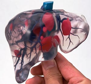
UCSD's 3D Printed Liver Cells Most Liver-like Yet from cysticfibrosis.com Learn vocabulary, terms and more with flashcards, games and other study tools. File edit view arrange extras help. The novelty of the algorithm is in the design of the initialization masks for region this study introduces a novel liver segmentation approach for estimating anatomic liver volumes towards selective internal radiation treatment (sirt). The liver resides in almost the entire length of the upper abdomen. Royalty free 3d model human liver damaged for download as ma, ztl, ma, fbx, and obj on turbosquid: Commonly used liver fibrosis models. You are able to do it by yourself with no wide range of dilemma. The cables are initially minimize by a specialized slicing machine and then they are printed on.
The liver region is further segmented using localized contouring.
File edit view arrange extras help. The negative effect of alcohol on the liver , the round diagram shows the reversible and irreversible effect of alcohol on the. The liver region is further segmented using localized contouring. Diagram of the liver and gall bladder showing the most. While the greatest portion sits in the right hypochondriac region, it extends past the epigastrium and over into the left hypochondriac region. In humans, it is located in the right upper quadrant of the abdomen, below the diaphragm. The liver resides in almost the entire length of the upper abdomen. Royalty free 3d model human liver damaged for download as ma, ztl, ma, fbx, and obj on turbosquid: Ordinarily, diagram of body liver harness assembly is created in keeping with geometric in addition to electrical requires. 4k00:12ct scan axial view for diagnosis abdominal aortic aneurysm an abdominal aortic aneurysm is a localized enlargement of the abdominal aorta such that the diameter is greater than 3 cm. It can be felt as a hardish vessels the liver receives approximately 30% of resting cardiac output and is therefore a very vascular organ. As in depth in this post, the answer depends upon two or three various factors. You are able to do it by yourself with no wide range of dilemma.